

:format(url)/cloudfront-us-east-1.images.arcpublishing.com/lescoopsdelinformation/L2F6OCLVY5CBBNQ32DX5S7TOBA.jpg)


The disease is really scary. But recent advances are important enough to restore hope and encourage early diagnosis of lung cancer. An already well-known institution in the field of cardiology, the Institut Arnaud Tazanke (IAT), in Saint-Laurent-du-Var, has created, under the impetus of its General Director, Michel Salvadori, a global offer of care, inviting specialists in the field of diagnosis, treatment and follow-up of lung cancer. Meeting with the IAT chest unit medical team, made up of pulmonologists, radiologists, surgeons, anesthesiologists and resuscitators, and all members of the multidisciplinary consultative meeting (RCP) that will decide on patient care.
How are lung nodules monitored?
In several ways: either as part of a smoker’s examination for chronic diseases, such as COPD. Either completely at random, during a CT scan for another reason, or even during a heart surgery evaluation [l’établissement a une forte activité dans ce domaine, Ndlr].
the following?
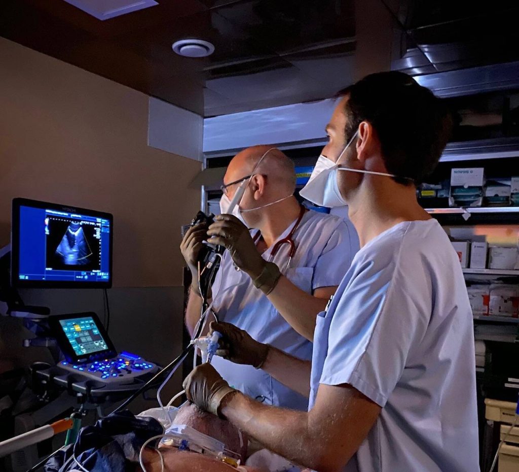
If these nodules are larger than 10 mm, additional investigations are performed without delay: a PET scan (a scan to assess the activity and extent of carcinomas), fibrosigmoidoscopy or bronchoscopy (depending on the location of the lesions) to determine the nature of the nodule and to assess the degree of spread of a potentially serious disease.
Aren’t these tests at risk of complications?
Removing the lesions under a scanner (and under light sedation) is very effective and has little risk. However, this risk is not zero and that is why these examinations are performed within the framework of a short hospital stay. It is necessary to secure all work in cooperation with the thoracic surgeons and resuscitators.
What about cases where the identified lesions are small?
In this case, a new examination is performed after four months, to assess the doubling time of the lesion. If it is short – which means that the lesion is developing rapidly – a puncture is made for analysis. Otherwise, we keep following.
When an abnormality in the lungs is identified, does it always indicate the presence of cancer?
No, in the vast majority of cases, these “abnormal” images correspond to benign lesions or scars from old infections such as tuberculosis or even concentrations of pollutants in the lungs. But imaging alone cannot distinguish between these benign abnormalities and neoplastic lesions.
When and how is the surgery done?
When histopathological examination confirms the presence of a neoplastic lesion – and intervention can be envisaged – the operation will involve excision of the tumor and adjacent lymph nodes. Depending on the extent of the lesion, the surgeon removes one of the five lobes of the lung (lobectomy) or exceptionally the entire lung. But the goal is to be as minimally invasive as possible, with the help of video-assisted surgery and robotic surgery. Thanks to the DaVinci Xi robot, the consequences of this complex surgery are much simpler; Postoperative pain and complications are greatly reduced.
It remains a major surgery that, to ensure its safety, requires the presence of a resuscitation service nearby.
alternatives?
On specific indications, validated by CPR, interventional radiologists can offer innovative radiofrequency tumor ablation procedures.
1. The team includes Dr. Olivier Castelnau, respiratory oncologist, Dr. Guy Boyer, interventional pulmonologist, Dr. Sebastian Novellas, interventional radiologist, Dr. Marc-Paul Francisci and Daniel Pope, thoracic surgeons, and Dr. Ghislaine Velotini, resuscitation specialist.

“Music guru. Incurable web practitioner. Thinker. Lifelong zombie junkie. Tv buff. Typical organizer. Evil beer scholar.”
More Stories
Sophie Adino officially receives her “wings” and will be able to fly in space by 2030
Plants grow mostly in the afternoon
Why should we not confuse academic freedom with the autonomy of science?